View the
Arrhythmia Recognition Webcast Series
|
 |
Practice ECG 4
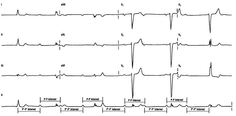
Don't panic! Break it down into its components as we've shown you before. Let's start off by looking only at the P waves. Use your calipers and mark the distance between what you know are two adjacent P waves. The best ones are between the fourth and fifth beats. There is a P wave right before the fourth QRS complex and one right after the T wave (the second hump). Now, place the calipers on the top of both of these P waves. Let's call this the P-P interval. Walk the calipers back and forth to find the other P waves. Do they map? Not exactly. Move your calipers to some other Ps, and you will note that every second interval matches this distance. The other P-P interval in this ECG-let's call it P'-P'-is slightly wider and also occurs every second time. To find this interval, place your calipers on the P wave right after the T of the fourth beat and on the P immediately in front of the fifth QRS complex. The P waves are mathematically linked but grouped separately. Forget about the name of the rhythm for now. We just threw this in to get you used to working with ECGs that are difficult. For advanced readers, this is ventriculophasic third-degree heart block, a rhythm in which any two Ps that have a QRS complex between them will be closer together (P-P) than those Ps that do not (P'-P').
There is always confusion around the terms third-degree heart block and AV dissociation. Dr. Chou makes a clear distinction in his book (see Additional Readings section): "The term complete AV block is used when the atrial rate is faster than the ventricular rate, whereas the reverse is true in AV dissociation. In AV block, there is a failure of impulse conduction even though the ventricles are receptive. In AV dissociation, there is an increase in the automaticity of the subsidiary pacemaker, which renders the ventricles functionally refractory to the slower atrial impulses." These are the definitions we have used in this book.
« Back to All
|